

Put simply, a tumour is a mass of the body’s own cells that multiplies. The word comes from Latin and roughly means ‘growth’ or ‘swelling’. Generally speaking, one could simply refer to skin tumours as skin cancer. Skin tumours can generally be classified as either benign or malignant, depending on their biological behaviour.
Skin tumours occur from time to time in chameleons. Studies indicate that skin tumours account for up to 10 per cent of all tumours in chameleons, and that malignant skin tumours are slightly more common than benign ones.1Heckers, K.O., Dietz, J., Marschang, R.E. (2015): Skin tumours in chameleons. Proceedings of the 44th Conference of the Working Group on Amphibian and Reptile Diseases, Gera: 95. The following overview presents a small selection of the skin tumours found in chameleons.
Papilloma
Since the 2000s, papillomas have been appearing with increasing frequency in chameleons kept in terrariums. They have a very distinctive, scaly appearance. In most cases, a primary papilloma first develops on the chameleon’s torso, after which further papillomas spread across the entire body over the following months.2Heckers, K.O., Dietz, J., Marschang, R.E. (2015): Papillomatosis in chameleons. Proceedings of the 44th Conference of the Working Group on Amphibian and Reptile Diseases, Gera: 87. Papillomas are found in many other reptiles and mammals.
Chromatophoroma
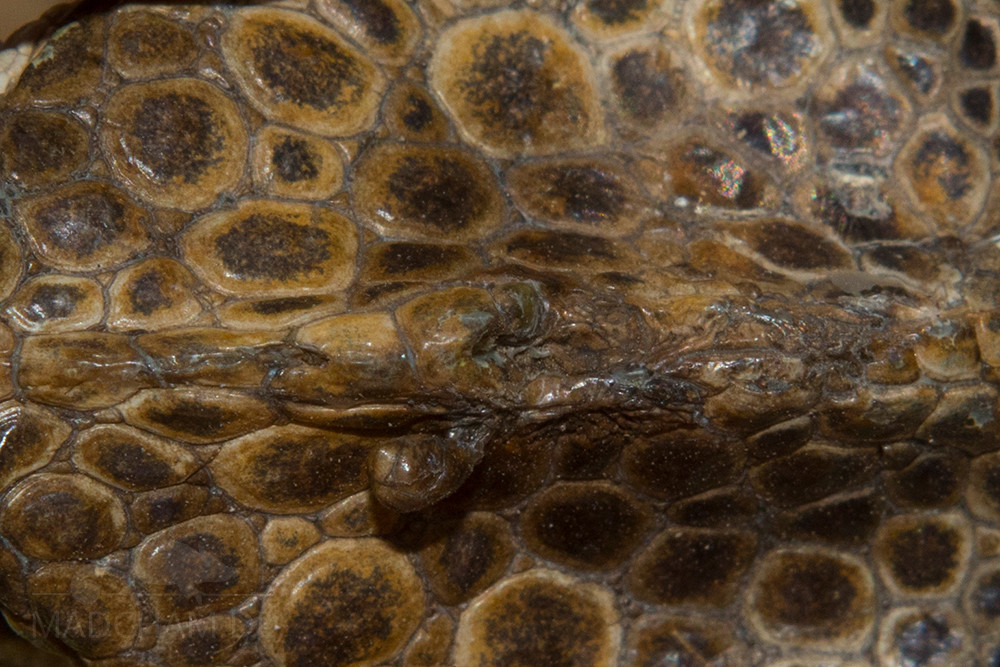
Skin tumour on the casque of a male Furcifer verrucosus
The so-called chromatophores, specialised skin cells, are responsible for the colour change in chameleons. They are arranged in layers beneath the outermost layer of skin, in the dermis. A tumour of these pigment-producing cells is called a chromatophoroma. Most chromatophoromas occur singly and are found on the head or chest of the chameleon.3Heckers, K.O., Aupperle, H., Schmidt, V., Pees, M. (2011): Melanophoromas and Iridophoromas in Reptiles. Journal of Comparative Pathology 146 (2): 1-11. Download In most cases, a histological examination in the laboratory can determine which type of chromatophore the tumour cells belong to. Chromatophoromas are then further classified into iridophoromas, melanophoromas and xanthophoromas.
Iridophoroma
An iridophoroma is a malignant tumour of the iridophores in a chameleon’s skin. To date, iridophoromas in chameleons have mainly been detected on the lateral body wall and the limbs.4Bronson, E., Pereira, M., Sanchez, C., Murray, S. (2006): Iridophoroma in a Veiled Chameleon, Chamaeleo calyptratus. Journal of Herpetological Medicine and Surgery 16(2): 58-60. 5Tong, L.J, Ong, W., Hulst, F., Tobias, G., Herrin, K.V., Vogelnest, L. (2018): Clinical, diagnostic, and pathological features of two cases of metastatic iridophoroma in a veiled chameleon (Chamaeleo calyptratus) and a red-barred dragon (Ctenophorus vadnappa). Journal of Exotic Pet Medicine 27: 53-60. 6Garner, M.M., Hernandez-Divers, S.M., Raymond, J.T. (2004): Reptile neoplasia: A retrospective study of case submissions to a specialty diagnostic service. Veterinary Clinic of North American Exotic Animal Practice 7(3): 653-671. They can spread rapidly throughout almost all of a chameleon’s body tissues.7Heckers, K.O., Aupperle, H., Schmidt, V., Pees, M. (2011): Melanophoromas and Iridophoromas in Reptiles. Journal of Comparative Pathology 146 (2): 1–11. Download
Melanophoroma
A melanophoroma is a tumour of the melanin-producing cells in a chameleon’s skin. These cells are actually responsible for the dark blue and black colours in chameleons. In mammals, this tumour is known as a melanoma. Melanophoromas occur more frequently (though not exclusively) on the back and head, i.e. mainly in the upper part of the body. Melanophoromas in chameleons are usually malignant and can spread to almost all tissues in the body.8Heckers, K.O., Aupperle, H., Schmidt, V., Pees, M. (2011): Melanophoromas and Iridophoromas in Reptiles. Journal of Comparative Pathology 146 (2): 1-11. Download 9Garner, M.M., Hernandez-Divers, S.M., Raymond, J.T. (2004): Reptile neoplasia: A retrospective study of case submissions to a specialty diagnostic service. Veterinary Clinic of North American Exotic Animal Practice 7(3): 653-671.
Xantophoroma
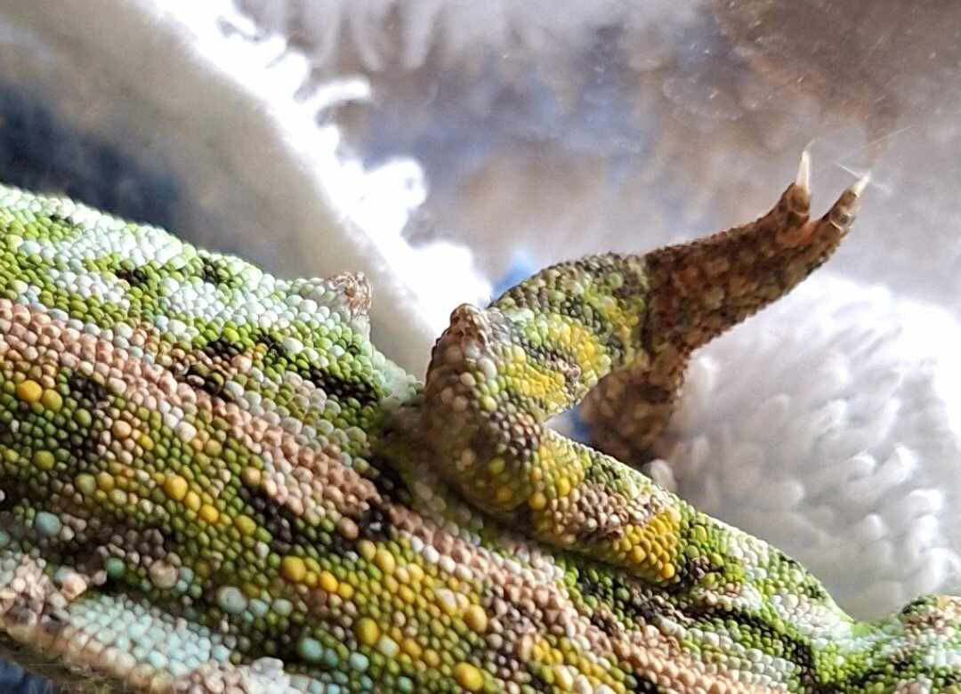
They look exactly like skin tumours, but histological examination revealed that these Furcifer lateralis specimens were in fact fungal granulomas.
Xantophoromas develop from xanthophorae. These are skin cells responsible for the yellow and orange colours in a chameleon’s skin. There are only a few case reports of xantophoromas in chameleons.10Garner, M.M., Hernandez-Divers, S.M., Raymond, J.T. (2004): Reptile neoplasia: A retrospective study of case submissions to a specialty diagnostic service. Veterinary Clinic of North American Exotic Animal Practice 7(3): 653-671. All of them developed metastases in various internal organs.
Squamous cell carcinoma
Squamous cell carcinoma originates in the skin’s epithelial and glandular tissue. It appears to be relatively common in chameleons and can occur anywhere on the body. Common sites include the head, the sides of the body and the limbs11Meyer, J., Kolodziejek, J., Häbich, A.-C., Richter, B. (2018): Multicentric squamous cell tumors in panther chameleons (Furcifer pardalis). Journal of Exotic Pet Medicine 29: 166-172., but the eyelid skin may also be affected.12Abou-Madi, N., Kern, T.J. (2002): Squamous cell carcinoma associated with a periorbital mass in a veiled chameleon (Chamaeleo calyptratus). Veterinary Ophthalmology 5(3): 217-220.. In most chameleon patients, several squamous cell carcinomas occur either simultaneously or in succession, at different sites.13Meyer, J., Kolodziejek, J., Häbich, A.-C., Richter, B. (2018): Multicentric squamous cell tumors in panther chameleons (Furcifer pardalis). Journal of Exotic Pet Medicine 29: 166-172. Squamous cell carcinoma is always malignant and tends to recur. Furthermore, squamous cell carcinoma can spread. Lung metastases are the most common.14Meyer, J., Kolodziejek, J., Häbich, A.-C., Richter, B. (2018): Multicentric squamous cell tumors in panther chameleons (Furcifer pardalis). Journal of Exotic Pet Medicine 29: 166-172.
Keratoacanthoma
This tumour, which is usually benign, develops from the skin’s keratinocytes – the cells that also form the scales on a chameleon’s skin. In chameleons, keratoacanthomas are usually found on the trunk.15Heckers, K.O., Dietz, J., Marschang, R.E. (2015): Hauttumore bei Chamäleons. Tagungsband der 44. Tagung der AG Amphibien- und Reptilienkrankheiten, Gera: 95.
How can you recognise a skin tumour?
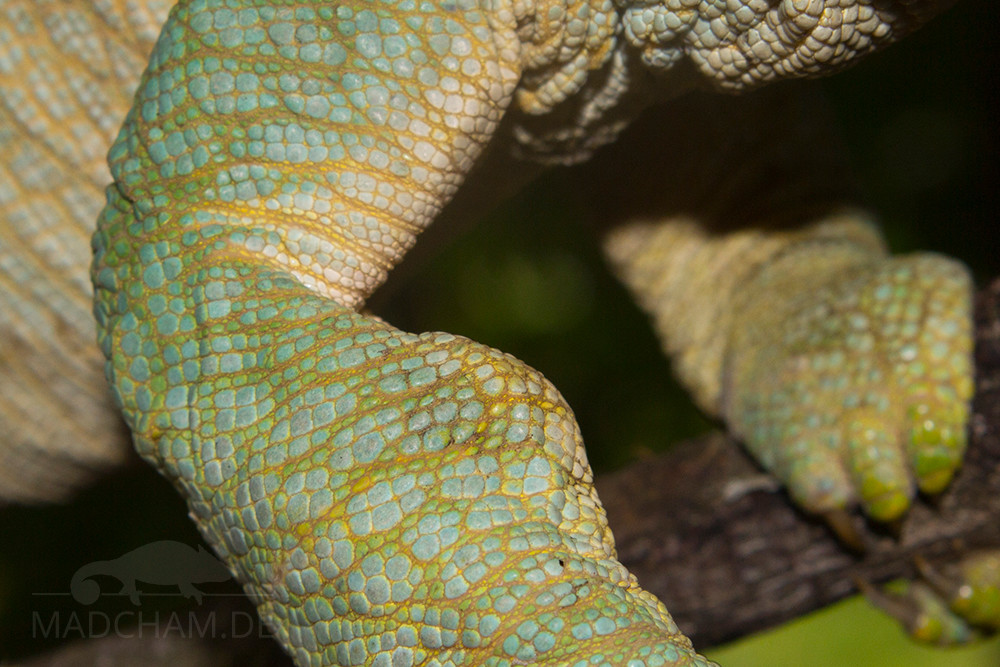
This swelling in the skin on the forearm of a Parsons’ chameleon could also be an abscess.
A skin tumour usually presents as a small raised area on the skin at first. Depending on the type of tumour, it may grow rather slowly or very quickly. With some skin tumours, the skin may change colour, for example turning black or greyish. The skin often loses its scaly texture. As a result, flakes of skin tend to stick to the area. But be careful! A lump on the skin is not necessarily a skin tumour. An abscess, necrosis (dead skin) or a fungal granuloma can also look exactly the same on the outside. It is therefore very important to have a diagnosis carried out by a vet specialising in reptiles in order to distinguish between them.
Causes
Skin tumours can have a wide variety of causes. Unfortunately, the cause of any skin tumour in chameleons has not yet been established. Given the increased incidence of chromatophoromas in chameleons in recent years, a link with artificial UV-B radiation is currently being discussed.16Meyer, J., Kolodziejek, J., Häbich, A.-C., Richter, B. (2018): Multicentric squamous cell tumors in panther chameleons (Furcifer pardalis). Journal of Exotic Pet Medicine 29: 166-172. 17Heckers, K.O., Aupperle, H., Schmidt, V., Pees, M. (2011): Melanophoromas and Iridophoromas in Reptiles. Journal of Comparative Pathology 146 (2): 1-11. Download Pet shops sell a wide range of unsuitable UV-B lamps, which either emit radiation outside the UV-B spectrum suitable for chameleons or, if fitted incorrectly – too close to or even inside the terrarium – can pose a hazard.
Viruses are also thought to be a possible cause of papillomas, as it has been shown that papillomas in other reptiles are caused by herpes, papilloma or iridoviruses. However, herpesviruses (chamHV) have so far been noted in chameleons primarily as the cause of scaly lesions on the lips.18Hellebuyck, T., Couck, L., Ducatelle, R., Van den Broeck, W., Marchang, R.E. (2021): Cheilitis associated with a novel Herpesvirus in two panther chameleons (Furcifer pardalis). Journal of Comparative Pathology 182: 58-66. However, there is as yet no evidence that herpesviruses – or indeed any other viruses – cause papillomas in chameleons.19Heckers, K.O., Dietz, J., Marschang, R.E. (2015): Papillomatose bei Chamäleons. Tagungsband der 44. Tagung der AG Amphibien- und Reptilienkrankheiten, Gera: 87.
It is also thought that advanced age and immunosuppression may generally contribute to tumour growth.
Diagnosis and treatment
Skin tumours should always be shown to a vet specialising in reptiles at an early stage. The smaller the tumour, the easier it is to remove surgically under anaesthesia. Furthermore, with malignant skin tumours, the likelihood of the tumour spreading to other organs, such as the liver or lungs, increases over time. In such cases, surgery may come too late. Before an operation, a blood test is usually carried out to assess the patient’s suitability for anaesthesia, and X-rays and/or an ultrasound scan may also be performed to detect any metastases.
If the skin tumour is in a very awkward location or is very large, the vet may first take a biopsy – that is, a tissue sample. A histopathological examination of the removed tissue in the laboratory will then reveal the nature of the tumour. If the entire tumour has been excised, the histological examination will also determine whether all the tumour tissue has been completely removed – in other words, whether healthy skin cells can be found around the tumour.This determines both the prognosis for the healing of the surgical wound and for the chameleon’s survival. Furthermore, the exact type of tumour indicates whether metastases (spread) or recurrences (the tumour coming back) are likely or not. If surgery is not possible, there are isolated case reports of successful cryotherapy for skin tumours. This involves treating the tumour cells with cold. However, for most skin tumours in chameleons, the most promising approach to date is surgery.
Literatur zu diesem Thema
- 1Heckers, K.O., Dietz, J., Marschang, R.E. (2015): Skin tumours in chameleons. Proceedings of the 44th Conference of the Working Group on Amphibian and Reptile Diseases, Gera: 95. ↩︎
- 2Heckers, K.O., Dietz, J., Marschang, R.E. (2015): Papillomatosis in chameleons. Proceedings of the 44th Conference of the Working Group on Amphibian and Reptile Diseases, Gera: 87. ↩︎
- 3
- 4Bronson, E., Pereira, M., Sanchez, C., Murray, S. (2006): Iridophoroma in a Veiled Chameleon, Chamaeleo calyptratus. Journal of Herpetological Medicine and Surgery 16(2): 58-60. ↩︎
- 5Tong, L.J, Ong, W., Hulst, F., Tobias, G., Herrin, K.V., Vogelnest, L. (2018): Clinical, diagnostic, and pathological features of two cases of metastatic iridophoroma in a veiled chameleon (Chamaeleo calyptratus) and a red-barred dragon (Ctenophorus vadnappa). Journal of Exotic Pet Medicine 27: 53-60. ↩︎
- 6Garner, M.M., Hernandez-Divers, S.M., Raymond, J.T. (2004): Reptile neoplasia: A retrospective study of case submissions to a specialty diagnostic service. Veterinary Clinic of North American Exotic Animal Practice 7(3): 653-671. ↩︎
- 7Heckers, K.O., Aupperle, H., Schmidt, V., Pees, M. (2011): Melanophoromas and Iridophoromas in Reptiles. Journal of Comparative Pathology 146 (2): 1–11. Download ↩︎
- 8
- 9Garner, M.M., Hernandez-Divers, S.M., Raymond, J.T. (2004): Reptile neoplasia: A retrospective study of case submissions to a specialty diagnostic service. Veterinary Clinic of North American Exotic Animal Practice 7(3): 653-671. ↩︎
- 10Garner, M.M., Hernandez-Divers, S.M., Raymond, J.T. (2004): Reptile neoplasia: A retrospective study of case submissions to a specialty diagnostic service. Veterinary Clinic of North American Exotic Animal Practice 7(3): 653-671. ↩︎
- 11Meyer, J., Kolodziejek, J., Häbich, A.-C., Richter, B. (2018): Multicentric squamous cell tumors in panther chameleons (Furcifer pardalis). Journal of Exotic Pet Medicine 29: 166-172. ↩︎
- 12Abou-Madi, N., Kern, T.J. (2002): Squamous cell carcinoma associated with a periorbital mass in a veiled chameleon (Chamaeleo calyptratus). Veterinary Ophthalmology 5(3): 217-220. ↩︎
- 13Meyer, J., Kolodziejek, J., Häbich, A.-C., Richter, B. (2018): Multicentric squamous cell tumors in panther chameleons (Furcifer pardalis). Journal of Exotic Pet Medicine 29: 166-172. ↩︎
- 14Meyer, J., Kolodziejek, J., Häbich, A.-C., Richter, B. (2018): Multicentric squamous cell tumors in panther chameleons (Furcifer pardalis). Journal of Exotic Pet Medicine 29: 166-172. ↩︎
- 15Heckers, K.O., Dietz, J., Marschang, R.E. (2015): Hauttumore bei Chamäleons. Tagungsband der 44. Tagung der AG Amphibien- und Reptilienkrankheiten, Gera: 95. ↩︎
- 16Meyer, J., Kolodziejek, J., Häbich, A.-C., Richter, B. (2018): Multicentric squamous cell tumors in panther chameleons (Furcifer pardalis). Journal of Exotic Pet Medicine 29: 166-172. ↩︎
- 17
- 18Hellebuyck, T., Couck, L., Ducatelle, R., Van den Broeck, W., Marchang, R.E. (2021): Cheilitis associated with a novel Herpesvirus in two panther chameleons (Furcifer pardalis). Journal of Comparative Pathology 182: 58-66. ↩︎
- 19Heckers, K.O., Dietz, J., Marschang, R.E. (2015): Papillomatose bei Chamäleons. Tagungsband der 44. Tagung der AG Amphibien- und Reptilienkrankheiten, Gera: 87. ↩︎
Das könnte dir auch gefallen


Follow us on Instagram

Most popular articles


Your travel specialist for Madagascar