
Parasites are a very common problem in chameleons, particularly in wild-caught animals and those captive-bred in large breeding facilities. Generally speaking, a distinction is made between endoparasites and ectoparasites. Endoparasites are those that colonise the inside of a chameleon. Ectoparasites, on the other hand, are found only on the chameleon’s skin. Parasites vary in how ‘disease-causing’ they are for chameleons: some are very harmful even with a minor infestation, whilst others remain harmless for a long time.
This article aims to provide an overview of the most common parasites found in chameleons, including how they are transmitted, their life cycles and how they can be detected. No treatment options are recommended here, as both the diagnosis and treatment of a parasitic infestation should always be discussed with a vet specialising in reptiles.
Inhaltsverzeichnis
How can you tell if a chameleon has parasites?
With ectoparasites, it’s quite simple: you can see them with the naked eye. Small, red bumps in the armpits or a large tick on the nose. With endoparasites, unfortunately, it’s not that simple. Most parasite eggs aren’t visible in the droppings at all. And if whole worms are already being passed, the infection is usually already very advanced. You shouldn’t let it get that far in the first place!
Almost all of the endoparasites presented here can be detected at an early stage through faecal examinations at the vet’s. But be careful: if nothing is found in the droppings, this does not automatically mean that the chameleon is free of parasites! Many stages of the parasite’s life cycle are not constantly excreted in the droppings. A single faecal sample is therefore not sufficient to rule out a parasite infestation. If you have a chameleon in quarantine, at least two, or better still three, faecal examinations are advisable to rule out or confirm a parasitic infection.
Endoparasites
Coccidia
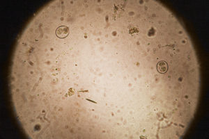
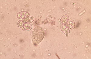
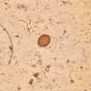
Oocysts of Eimeria vencesi in a native faecal smear of Furcifer pardalis, magnified 100 times
The bane of chameleon keeping: one in two large-scale breeding operations has problems with coccidia. Coccidia are single-celled organisms. Currently, over 20 different species are known to be capable of parasitising chameleons. The most important genera are Choleoeimeria, Eimeria and Isospora. Contrary to rumours to the contrary, coccidia are never part of a chameleon’s normal intestinal flora.
Life cycle
The infectious stages of coccidia are known as sporulated oocysts. Oocysts are present in large quantities in the faeces of infected chameleons. So when an infected chameleon defecates, a large number of coccidian oocysts, invisible to the human eye, end up in the surrounding environment. They can stick to branches that the chameleon has rubbed its cloaca against, to leaves or soil onto which the faeces have fallen. Insects walking over the faeces can carry the oocysts further afield. The next chameleon becomes infected by these oocysts by eating such an insect, accidentally ingesting a leaf containing oocysts, or licking a branch. A chameleon can even become infected at hatching: specifically, if the mother had coccidia oocysts in her cloaca whilst laying eggs. The oocysts end up on the eggshell and later, during hatching, on the juveniles. Infection via contaminated drinking water or objects to which oocysts are attached is also possible.
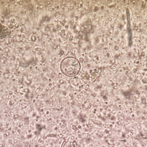
Oocysts of Choleoeimeria brookesiae in a native faecal smear of a Brookesia stumpffi, magnified 400 times
The coccidian oocysts are eventually swallowed and enter the chameleon’s intestine. There, they release specific parasitic stages which penetrate the intestinal mucosa and destroy it. Choleoeimeria prefer to settle in the gallbladder, where they cause damage. Through a relatively complex cycle, the coccidia develop into specific parasitic stages that are capable of reproducing. The oocysts are then excreted via the chameleon’s intestines in the faeces. This marks the start of the parasite’s cycle all over again.
In chameleons, it takes approximately three weeks for Isospora and five weeks for Choleoeimeria from the ingestion of coccidian oocysts to the excretion of new oocysts.
What problems do chameleons get from coccidia?
Coccidia are found in chameleons both in the wild and in captive environments. The intestinal lining damaged by coccidia often ceases to function properly. This frequently leads to diarrhoea. The chameleon becomes dehydrated, even though it drinks normally or even more than usual. Choleoemeria can lead to an enlarged gallbladder and a blocked bile duct, both of which are painful. In the long term, an undetected coccidian infection can lead to severe kidney disease.
Coccidia are particularly dangerous for juveniles, especially hatchlings. They usually die from coccidia within a short time. Entire clutches can be affected, with the juveniles dying one by one within a few weeks.
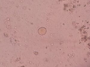
Oocysts of Choleoeimeria ssp. trematode egg in a native faecal smear of a chameleon, magnified 400 times
In adult chameleons, certain species of coccidia can sometimes cause an asymptomatic infection. The animals then show no signs of illness. Sometimes, the only noticeable symptom is weight loss. However, under
In adult chameleons, certain species of coccidia can sometimes cause an asymptomatic infection. The animals then show no signs of illness. Sometimes, the only noticeable symptom is weight loss. However, under stressful conditions such as mating, relocation or suboptimal husbandry conditions, coccidia can multiply particularly rapidly. In such cases, they can also lead to serious illness in these chameleons. As every chameleon experiences situations during its lifetime that can lead to an increase in coccidian infection, coccidia should never be regarded as harmless.
Incidentally, humans do not themselves suffer any problems from chameleon coccidia.
How do you get rid of coccidia?
The chameleon can be treated with medication administered orally. The medication must be carefully measured out by the vet, but is generally well tolerated.
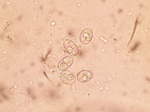
Oocyst of Isospora ssp. in a native faecal smear of a Furcifer pardalis, magnified 800 times
Coccidia oocysts are, unfortunately, extremely hardy. Under favourable conditions, they remain infectious for over a year. Neither cold nor heat affects them. Heat only kills coccidia if temperatures exceed 60°C for several minutes. Coccidia are resistant to most chemical disinfectants. Sterilium, which is used for hand disinfection in humans, and many of the disinfectants for terrariums commonly available in pet shops are, unfortunately, useless against coccidia. Chlorocresol is effective, but due to its harmful effects on human health, it should only be used in consultation with a vet. The high resistance to ‘simpler’ disinfection methods and a lack of quarantine are likely the reasons why coccidia are, unfortunately, very widespread in chameleon keeping.
Amoeba
Amoebae are a very large group of single-celled organisms. Only a very few are pathogenic to chameleons. The best-known of these is Entamoeba invadens. It is very rare in chameleons, but when it does occur, it is fatal. Amoebae are more common in other reptiles, such as tortoises.
Life cycle
The reproductive stages of amoebae are known as cysts. These cysts are present in large numbers in the faeces of infected chameleons. Infected chameleons defecate into their surroundings, causing branches, insects or simply the substrate to come into contact with the cysts. The next chameleon becomes infected by ingesting these cysts from the environment, for example during a tongue test or when it captures an insect that has previously walked over the faeces. The cysts, which are invisible to the human eye, are swallowed and eventually end up in the chameleon’s large intestine. There, they penetrate the intestinal wall. Via blood vessels, they reach the liver, kidneys and other organs. At the same time, new cysts are released into the intestine, which the chameleon then excretes in its faeces.
What problems do amoebae cause for the chameleon?
Cyst of Entamoeba ssp. im a native faecal smear, magnified 1600 times; CC BY-SA 4.0 Ajay Kumar Chaurasiya
The condition as a whole is known as amoebiasis. It is characterised primarily by severe, bloody intestinal inflammation accompanied by diarrhoea, weight loss and dehydration. In some cases, parts of the intestine may even begin to die off gradually. Amoebae also cause severe inflammation in other organs, such as the kidneys; in advanced stages, this ultimately leads to organ failure and, consequently, the death of the chameleon.
How do you get rid of amoebas?
The chameleon can be treated with medication prescribed by a vet, which is administered into its mouth. The medication is generally well tolerated.
Amoeba cysts can survive in the soil for at least eight days and can be spread via feed insects or by clinging to objects. They are highly infectious, meaning they spread extremely easily. In the terrarium, the cysts are mainly eliminated by heat. A temperature of over 60°C must be maintained for a good ten minutes.
As Entamoeba invadens develops optimally at a body temperature of 27–29°C, only reptiles are affected by these amoebae. Humans can contract amoebic dysentery (severe diarrhoea) from another amoeba called Entamoeba histolytica, but not from Entamoeba invadens. This parasite is therefore dangerous for chameleons and other reptiles, but not for humans.
Ciliates

Nyctotherus ssp. cyst in a native faecal smear, magnified 400 times
Ciliates are commonly found in the faeces of many chameleons. Many species are harmless to chameleons. Only under poor husbandry conditions or in the presence of pre-existing conditions can they occasionally cause problems in the event of a mass infestation. Among the better-known ciliates are the genera Nyctotherus and Balantidium, both of which use cysts as a reproductive stage.
Life cycle
The chameleon ingests infectious cysts from the faeces of another chameleon. It swallows the cysts, which then enter its intestines. In the intestines, the ciliates multiply and form cysts of their own. These cysts are then excreted in the chameleon’s faeces.
What problems do chameleons get from ciliates?
Generally speaking, none at all. However, if the chameleon is already suffering from other conditions, it may be advisable to treat any ciliates present as well. The treating vet will provide appropriate advice.
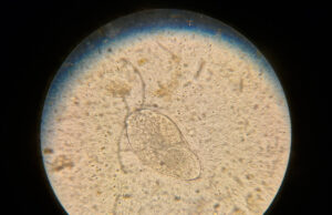
Live ciliated protozoa in a native faecal smear, magnified 400 times
How do you get rid of ciliates?
If necessary, the chameleon can be treated with medication administered orally after being prescribed by a vet. It is not usually necessary to treat the surroundings.
Flagellates
Flagellates is an umbrella term for a whole range of motile single-celled organisms. They are characterised by long cellular appendages that enable them to move – the flagella, from which they take their name. Whether flagellates cause illness in a chameleon depends on the species of flagellate, as well as the chameleon’s age and state of health. Well-known genera of flagellates found in chameleons include Leptomonas, Monocercomonas, Hexamita and Trichomonas.
Life cycle
Trichomonas ssp. in native faecal smear, magnified 400 times
In infected chameleons, flagellates are present in large numbers in faeces and urate. The chameleon’s excrement, containing live flagellates and cysts, ends up on the ground. The cysts stick to leaves, branches, plants or moss. If another chameleon ingests cysts via a prey animal, it becomes infected with the flagellates. The chameleon swallows the cysts, causing them to end up in the intestine. There, they develop into live flagellates, which in turn produce more cysts. These are then excreted in the faeces.
Infection with live flagellates is primarily possible via prey insects and is known to occur with the genus Leptomonas. Some flagellates can infect not only the gut but also other organs such as the liver, kidneys or lungs.
What problems do chameleons get from flagellates?
Countless Leptomonas ssp. in a fresh faecal smear, magnified 400 times
Flagellates are found in captive-bred chameleons as well as in wild-caught specimens and those living in the wild. An infestation involving only a few flagellates is often overlooked at first, as the chameleon shows no signs of illness. With flagellate species that are not particularly pathogenic, problems rarely arise even later on. Hexamita, on the other hand, relatively frequently cause kidney damage. Leptomonas often lead to bloody intestinal inflammation accompanied by foul-smelling diarrhoea.
How do you get rid of flagellates?
An infestation with Leptomonas or Hexamita in chameleons can be treated with medication prescribed by a vet, which is administered orally. The medication is generally well tolerated.
Following a flagellate infestation, the terrarium can be heat-treated for disinfection. The flagellates themselves dry out very quickly. The cysts, however, remain infectious for a long time even under adverse conditions. A temperature of over 60°C reliably kills the cysts.
Trematodes (flukes)
Flukes are parasitic flatworms. They usually have a flat body and the suction cups on their underside. Around 30 different species of flukes are currently known to infect chameleons. The best-known genera are Laureriella, Paradistomoides and Plagiorchis.
Life cycle
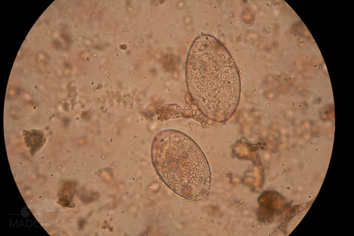
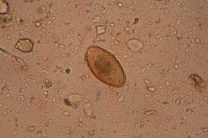
Two trematode eggs under the microscope in a native faecal smear, magnified 400 times
Trematode eggs are excreted by infected chameleons in their faeces, and less commonly in their urate. If the faeces land in a damp environment, such as a puddle or a pond, the eggs hatch into ciliated larvae (miracidia). The cystic larvae then burrow into the tissue of their intermediate host, often snails. The intermediate host, carrying the so-called tailed larvae (cercariae), is later eaten by the chameleon. Adult trematodes then develop in the chameleon’s intestine. These migrate to their preferred tissue. For most trematodes, this is the intestinal mucosa. In some species, however, it is also the oral cavity, the oesophagus, the stomach or even the gallbladder. There, they release eggs into the chameleon’s faeces. The next chameleon cannot become infected by the fluke eggs themselves, but only by the tail larvae within the intermediate host.
What problems do chameleons get from trematodes?
Flukes are found exclusively in wild chameleons or in wild-caught specimens intended for export. In captive-bred chameleons, flukes are virtually non-existent because the intermediate hosts required for their development are absent in the terrarium. Unfortunately, the fact that they occur only in the wild also means that little is known about the diseases that trematodes actually cause. It is assumed that trematodes are not particularly pathogenic and tend to cause localised inflammation in the gastrointestinal tract. However, there have also been a few cases of mass infestation in wild chameleons in Madagascar.
How do you get rid of flukes?
A fluke infection in a chameleon can be treated with a medication that is administered orally or injected under the skin. However, it is not well tolerated.
The terrarium does not need to be treated in the event of a fluke infection. As long as there are no intermediate hosts, such as snails, the infection cannot be transmitted to other chameleons.
Cestodes (tapeworms)
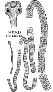
Schematic diagram of a tapeworm, graphic by Pearson Scott Foresman
Tapeworms are commonly found in many domestic animals, including dogs and cats. Like flukes, they belong to the phylum Platyhelminthes. At one end of their body, they have a hook-like structure with suction cups (scolex). The other end of the body is divided into many small segments, known as proglottids. Each proglottid is hermaphroditic, possessing both male and female reproductive organs. The genus Oochoristica, in particular, parasitises chameleons, among others.
Life cycle
Tapeworms live in the small intestine of chameleons, where they attach themselves to the intestinal walls using their suckers. The proglottids fertilise each other and are subsequently separated from the rest of the tapeworm’s body. They then leave the infected chameleon in its faeces. The faeces land on soil, moss or leaves. Every single proglottid is packed full of eggs. Six-hooked larvae hatch from these eggs, which must then be ingested by an intermediate host. In the intermediate host, the tapeworm larva embeds itself in the connective or muscle tissue in the form of a cyst (metacestode). If the intermediate host is then eaten by the chameleon, the metacestode passes through the stomach into the chameleon’s intestine. There it eventually develops into a tapeworm. A few species also colonise the gallbladder.
What problems do tapeworms cause in chameleons?
Tapeworms are found exclusively in wild chameleons or in wild-caught specimens intended for export. Chameleons bred in captivity generally do not contract tapeworms due to the lack of suitable intermediate hosts. Severe tapeworm infestation can lead to weight loss despite a good appetite. In rare cases, it can also cause intestinal inflammation accompanied by diarrhoea.
How do you get rid of tapeworms?
A tapeworm infestation in a chameleon can be treated with a medication that is administered orally or injected under the skin. However, it is not well tolerated.
The terrarium does not need to be treated in the event of a tapeworm infestation. As long as there are no intermediate hosts present, transmission to other chameleons cannot occur.
Nematodes (roundworms)
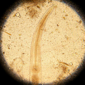
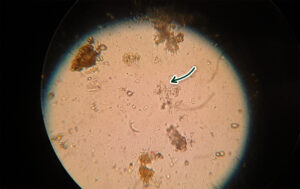
The anterior section of a roundworm in a fresh faecal smear from a chameleon, magnified 400 times
Nematodes look exactly as their name suggests. They are small, elongated and thin. There are more than 20,000 different species worldwide. Only a few of these are parasitic. Nematodes reproduce sexually. Males and females mate, after which the female produces eggs. In infected chameleons, these eggs are excreted in the The anterior section of a roundworm in a fresh faecal smear from a chameleon, magnified 400 times. A larva develops inside each egg. The various larval stages in roundworms are designated L1, L2, L3 and L4, with the pre-adult stage referred to as L5. Between each new stage, the larva undergoes moulting. The fifth larva, L5, eventually matures into an adult roundworm.
A notable feature of nematodes is their ability to enter hypobiosis. This term refers to a suspension of development at the third, fourth or fifth larval stage. During this ‘dormant phase’, the larvae remain in the tissue where they are currently located, waiting for a more favourable time to continue their development. Depending on exactly where in the chameleon’s body the larva is located during hypobiosis, it may be difficult or impossible to treat with medication. After hypobiosis, the larva can develop further without any problems and produce the next generation of nematodes. In the case of roundworms, it is therefore possible that no roundworm eggs can be found in a chameleon’s faeces, yet there are still larvae in hypobiosis within the body. If these are reactivated and continue to develop, the chameleon may once again excrete roundworms.
As there are so many different types of nematodes affecting chameleons, the following sections provide a more detailed overview of individual nematode species.
Nematodes: Rhabdiasidae (lungworms)
Rhabdiasidae are lungworms characterised primarily by a very interesting mode of transmission. According to current scientific understanding, it appears that each species of chameleon has its own specific Rhabdias species. It is therefore likely that there are significantly more lungworms than the 15 or so species currently known. Lungworms are found primarily in humid habitats such as rainforests.
Life cycle

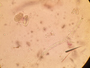
Rhabdias ssp. in a fresh faecal smear from a pygmy chameleon, magnified 400 times
Infected chameleons excrete infectious larvae in their faeces. These larvae actively emerge from the faeces and burrow into the skin of another chameleon that happens to be passing by. They enter the chameleon’s lungs via the bloodstream. Some species remain in the lungs and reproduce there. In other species, the larvae migrate along the windpipe into the chameleon’s mouth. There they are swallowed and thus reach the intestine. There, the larva develops into an adult lungworm. Only females exist, which produce eggs containing larvae through parthenogenesis without the presence of a male. The female lungworms effectively ‘clone’ themselves.
What problems do chameleons get from lungworms?
Lungworms are relatively common in the wild and in exported wild-caught animals. Leaf chameleons in particular seem to be severely affected. As the worms reside in the lungs and air sacs, affected chameleons will sooner or later develop breathing difficulties as a result of pneumonia. Increased mucus can be seen in the mouth. As a result of the severe pneumonia, the chameleon often loses weight.
How do you get rid of lungworms?
Rhabdias ssp. eggs and worm in a fresh faecal smear, magnified 100 times
In principle, lungworm infection in chameleons can be treated with drugs that are generally well tolerated. However, a major problem is that this treatment kills the worms inside the lungs and air sacs as well, not just those in the intestines. The dead lungworms cause severe inflammatory reactions, which in turn require treatment. It is therefore not uncommon for both lungworm infection and the treatment of lungworm infection in chameleons to unfortunately result in death. This is a very difficult situation. In many cases, thorough diagnostic testing can be carried out in advance by a vet to determine whether worms are present in the lungs and other body tissues, thereby assessing the risk.
As with many other parasites, the terrarium must be thoroughly cleaned and disinfected following a lungworm infestation before the chameleon can be returned to it. Heat of over 60°C for at least five minutes – preferably longer – will reliably kill the eggs.
Nematodes: Strongylidae (strongylids) and Molineoidae (threadworms)
Among the strongylids, the genus Kalicephalus is particularly well known, as it is always pathogenic to chameleons. The nematodes include the genus Oswaldocruzia, which has already been detected in several species of chameleon. Both families of parasites are so similar that they are discussed together here.
Life cycle
Egg containing larvae of Kalicephalus ssp. or Rhabdias ssp. in a native faecal smear from Brookesia stumpffi, magnified 400×
Infected chameleons excrete eggs containing larvae in their faeces. In the environment, the larvae develop into infectious larvae. These larvae are accidentally ingested by other chameleons whilst feeding, or they actively emerge from the faeces and burrow into the skin of other chameleons. They then travel to the lungs via the lymphatic system. From there, the fourth larva travels via the trachea into the oral cavity. The chameleon then swallows the larva, which enters the intestine. It is only there that it develops into an adult worm. Strongylids and nematodes burrow into the intestinal mucosa and lay eggs in the chameleon’s faeces.
What problems do chameleons face from strongylids or threadworms?
These worms cause problems, particularly in cases of mass infestation. The damaged intestinal walls become inflamed, leading to severe diarrhoea. Furthermore, chameleons frequently suffer from anaemia when infected with strongylids.
How do you get rid of strongyles and threadworms?
Strongylid infections in chameleons can be treated by a vet using very well-tolerated medication.
As with many other parasites, the terrarium must be thoroughly cleaned and disinfected following an infestation before the chameleon can be returned to it. Heat of over 60°C for at least five minutes – preferably longer – will reliably kill the eggs.
Please note: Strongylids are not particularly host-specific. This means they can also be transmitted to or acquired from other reptile species. In the event of a strongylid infestation, it is therefore advisable to check the entire reptile stock (and, unfortunately, to treat them if necessary).
Nematodes: Filarioidea (filariae)
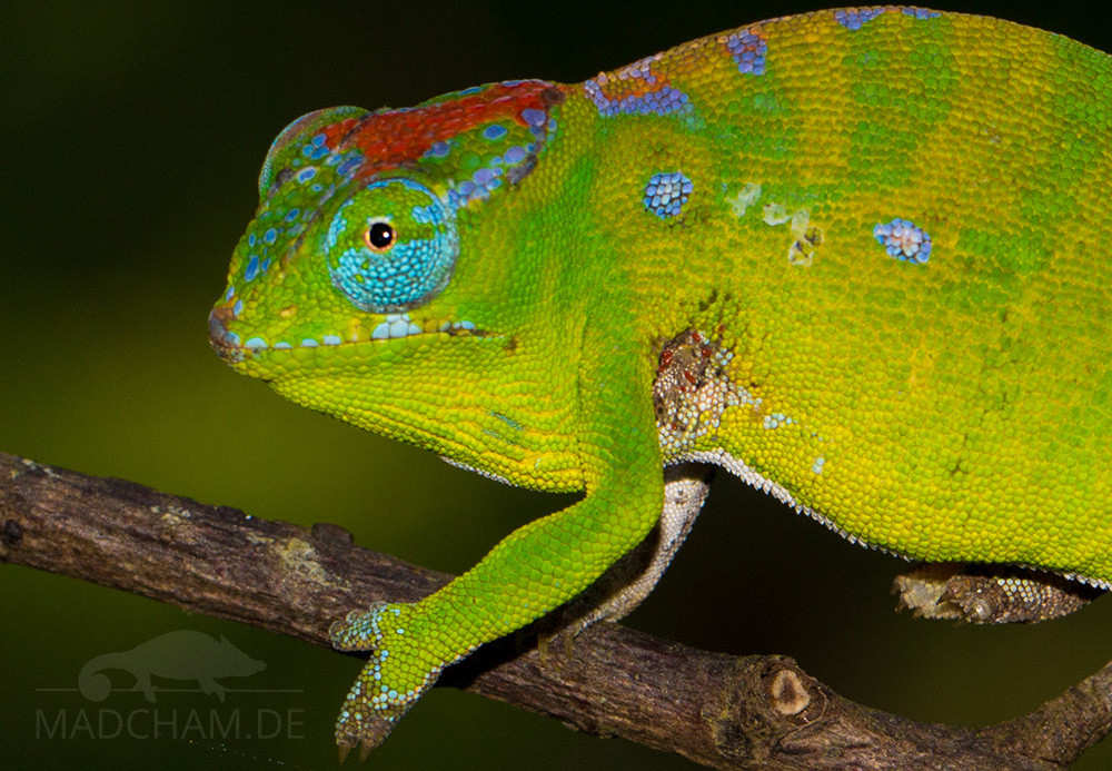
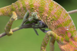
Macrofilaria or roundworms in the subcutaneous tissue of a young Furcifer pardalis in Akanin’ny Nofy
Filarial worms are slender nematodes that are relatively common in Madagascar and can grow from a few millimetres to eight centimetres in length. To date, only three species are known to infect chameleons: Foleyella brevicauda, Foleyella candezei and Foleyella furcata. In the subcutaneous tissue of an infected chameleon, the filariae can sometimes be seen as small, moving worms, which often seem to disappear again when light pressure is applied with a finger.
Please note: Filaria are detected via a blood smear. Unlike almost all other internal parasites, they cannot be found in the faeces.
Life cycle
The life cycle of filariae begins when a mosquito bites an infected chameleon and thereby ingests filariae. These develop further within the mosquito. If the same mosquito then bites another chameleon, it transmits the filariae. The first larval stage in the chameleon’s blood is called a microfilaria. These microfilariae travel through the chameleon’s bloodstream to various organs, where they continue to develop. Once fully grown, the parasites are known as macrofilaria. The macrofilaria eventually migrate to the coelomic cavity, the lungs or air sacs, or the subcutaneous tissue. They release microfilariae into the bloodstream, which can then be ingested by the next mosquito.
What problems does the chameleon get from filariae?
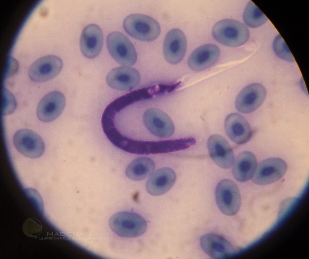
Microfilariae in a blood smear from Furcifer pardalis, magnified 400 times
Filariae are transmitted exclusively by certain species of mosquito, meaning that this nematode is found only in the wild and in exported wild-caught specimens. A minor infestation does not lead to disease. However, a mass infestation can result in what is known as filariasis. In doing so, microfilariae block blood vessels, causing the tissue that would normally be supplied by those vessels to die off. The migration of macrofilariae within the coelomic cavity can also lead to severe inflammation. In the worst-case scenario, this can prove fatal for the chameleon.
How do you get rid of filariae?
In theory, treating the chameleon itself would be sufficient to eliminate a filaria infection. Unfortunately, however, filariae are very difficult to treat with medication at the vet’s. The only medication known to be effective against these parasites is very poorly tolerated by chameleons and has, to date, consistently proved fatal.
Macrofilaria or roundworms in the subcutaneous tissue of a young Furcifer viridis in north-western Madagascar
As filariae always require a mosquito as an intermediate host, there is no need to disinfect the terrarium to prevent transmission of this parasite. A chameleon cannot become infected with filariae even if it were to share the same terrarium with an infected chameleon. However, it cannot be ruled out with absolute certainty that European, Asian or American mosquitoes might also transmit microfilariae from one chameleon to another. This could be important when keeping infected and uninfected chameleons together.
Nematodes: Ascaridida (roundworms)
Roundworms are found in almost all mammals and reptiles. When fully grown, they look like thick spaghetti and are therefore the ‘classic’ parasitic worm. In relation to their host’s body size, roundworms can reach impressive lengths. The human roundworm, for example, can grow up to 40 cm long! In chameleons, roundworms can grow up to 17 cm in length, which is quite impressive depending on the species of chameleon. Over 20 different species of roundworm have already been identified in various chameleon species, including, amongst others, the genera Hexametra and Orneoascaris. The Ascaridae family also includes the so-called Heterakidae, comprising the genera Africana, Heterakis, Strongyluris and Spinicauda.
Life cycle
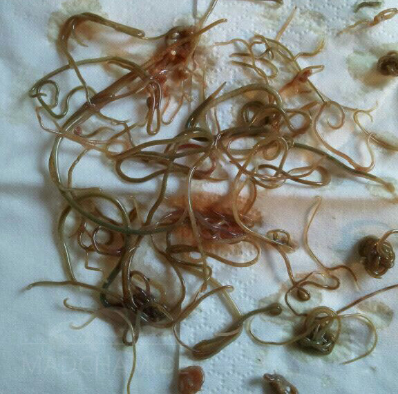
A vast number of roundworms from the coelomic cavity of a panther chameleon that died as a result of the severe infestation
Infected chameleons excrete large quantities of roundworm eggs in their faeces. The faeces fall onto leaves, branches and the ground. The roundworm eggs stick to whatever surface the faeces land on. And anything that has come into contact with the faeces – be it insects that have walked over them or a human hand cleaning up the terrarium – can carry the eggs further. The eggs contain the first larval stage of the roundworms. In the environment, the larvae in the eggs continue to develop into the infectious third larval stage.
Other chameleons then become infected by these eggs, which they carry around, when they accidentally take them into their mouths whilst performing tongue-testing behaviour or feeding. The roundworm eggs are swallowed and eventually end up in the small intestine. There, the larva hatches and develops into an adult roundworm, which lays eggs in the chameleon’s faeces.Roundworms also undertake migrations similar to those of filarial worms outside the small intestine. They migrate to the subcutaneous tissue, the liver, the fat bodies, the lungs and the air sacs. In some species of roundworm, which are particularly common in chameleons, both life cycles involving an intermediate host (or hosts) and those without an intermediate host are known to occur.
Egg of Spinicauda ssp. in a fresh faecal smear from Furcifer pardalis, magnified 400 times
As roundworm eggs are invisible to the naked eye but are usually present in large numbers in the vicinity of infected chameleons, they are often carried around unnoticed. In this way, the roundworm eggs are transferred from terrarium to terrarium, to new owners at pet fairs, and to new hosts via live feed containers, all without being detected by humans.
What problems do roundworms cause in chameleons?
At first, there are few noticeable signs. Although the chameleon eats well, it continues to lose weight. Roundworms can cause bloody ulcers, intestinal perforations and constipation in the gut due to a massive infestation. In the worst-case scenario – which, unfortunately, occurs regularly with roundworms in chameleons – the constipation leads to a fatal intestinal obstruction. Their migration outside the intestines can also lead to inflammation of the affected tissues. Roundworms in the lungs can even cause shortness of breath.
How do you get rid of pinworms?
The good news about roundworms: treating your chameleon at the vet involves a medication administered orally, which is simple and well tolerated. The bad news: if the infestation is detected too late and there is already a massive infestation, the dead worms can lead to intestinal blockage or inflammation of the organs.
Egg of Hexametra ssp. in a fresh faecal smear from Furcifer lateralis, magnified 400 times
Getting a terrarium back to a parasite-free state after a roundworm infestation through cleaning and disinfection is not quite so straightforward. The infectious eggs are extremely hardy and can survive for years in damp soil. Roundworm eggs are resistant to most chemical disinfectants. Unfortunately, Sterilium, which is used for hand disinfection in humans, or the disinfectants commonly available in pet shops for terrariums, are ineffective against nematode eggs. Ammonia or p-chloro-m-cresol should only be used in consultation with a vet, as they are harmful to human health.
Please note: Unfortunately, some species of roundworm are not host-specific. This means that, in addition to chameleons, other reptiles can also become infected by roundworm eggs. In the case of Hexametra angusticaecoides, for example, infections in various geckos and snakes have been reported. If a chameleon is infected with this roundworm species, it is therefore possible that other reptiles in the collection are also infected with the roundworms, and treatment of the entire animal stock may be necessary.
Nematodes: Oxyurida (pinworms)
Oxyurida are also known as pinworms. This genus of nematodes, which remain very small, is very common in reptiles. In chameleons, they are among the most common endoparasites, alongside coccidia. However, Oxyurida species are highly host-specific. This means that practically every reptile species has its own specific species of pinworms. Or to put it another way: the pinworms of turtles do not like chameleons or snakes, and vice versa. Among chameleons, the species known to date are Pharyngodon dimorpha, Thelandros meridionalis and Parapharyngodon kenyaensis.
Life cycle
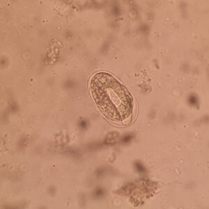
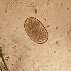
Oxyurida egg in a fresh faecal smear from Furcifer pardalis, magnified 100 times
Infected chameleons excrete needletail eggs in their faeces, which then stick to surfaces in the surrounding area. The eggs contain the first larval stage, which develops into the infectious third-instar larva in the environment. Other chameleons can then become infected from the eggs containing the infectious larvae if they ingest leaves bearing the eggs or insects that have walked over the faeces of an infected chameleon. The eggs are then swallowed along with the chameleon’s food and end up in its intestines. There, the pinworm larvae hatch from the eggs and develop into adult pinworms. The female pinworms then lay eggs of their own, which are passed in the chameleon’s faeces and begin a new life cycle.
The entire life cycle, from the eggs being ingested to their excretion, takes between four and six weeks.
What problems do chameleons get from pinworms?
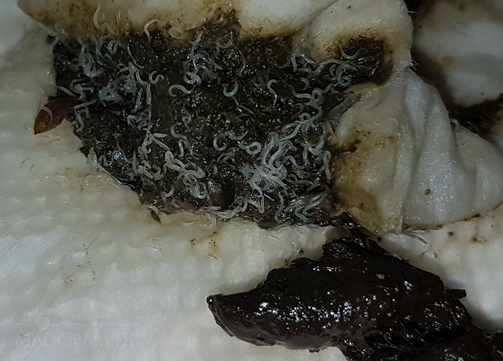
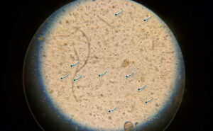
A massive infestation with pinworms, already visible to the naked eye in the faeces – this animal had been suffering from an unnoticed and, consequently, untreated infestation for years
Pinworms only cause illness in chameleons when the infestation is severe. They often go completely undetected until the first faecal examination. However, if the chameleon does fall ill at a later stage, the pinworms can multiply rapidly and lead to oxyuriasis, an infestation with clear signs of illness.
How do you get rid of pinworms?
A case of thrush in chameleons is very easy for a herp vet to treat. There are several medications that can be administered orally, all of which are very well tolerated. It is best to start treatment – including cleaning and disinfecting the terrarium – as soon as possible, even before the chameleon shows any symptoms of the condition.
The eggs are invisible to the naked eye. The eggs of pinworms remain infectious in the terrarium for months and are therefore particularly prone to being accidentally spread from one animal to another. As with many other parasites, the terrarium must be thoroughly cleaned and disinfected following an infestation before the chameleon can be returned. Heat exceeding 60°C for at least five minutes, preferably longer, will reliably kill the eggs.
Ectoparasites
Acari (mites and ticks)
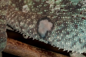
Mites in the armpit of a female Furcifer petteri in the Montagne d’Ambre
Mites and ticks belong to the arachnid family and develop from an egg into a larva. The larva develops into an adult parasite via a certain number of nymphal stages. The larvae have three pairs of legs, whilst the adults have four. In some species, a nymph hatches directly from the egg. Each stage of the life cycle is marked by a moult and a blood meal.
Mites
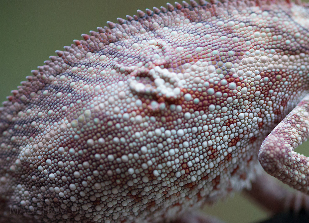
Mites are biting-sucking arachnids that are most commonly found around the eyes, in skin folds such as the armpits, and around a chameleon’s cloaca. Most mites are only one to two millimetres in size and bright red in colour. They can be easily spotted with the naked eye if you look closely. As mites feed on blood, a severe infestation can lead to anaemia in the chameleon.
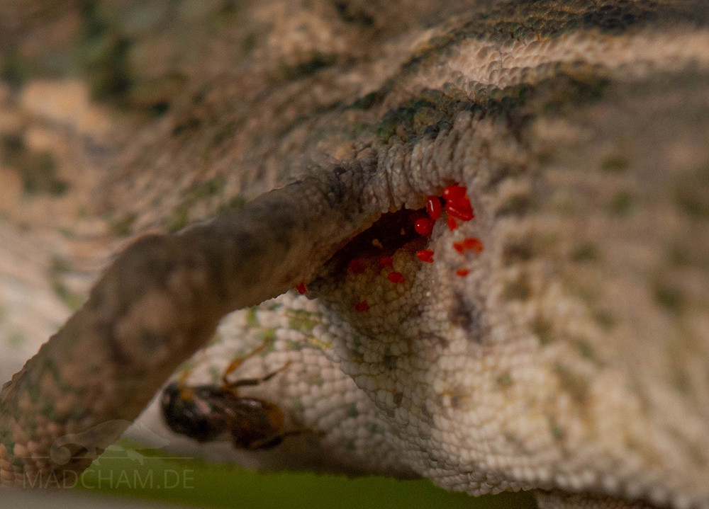
Mites in the armpit of a male Furcifer rhinoceratus in Ankarafantsika
Wild-caught and wild chameleons are particularly prone to mite infestations. In Madagascar, mites are quite common on all kinds of chameleons, especially during the rainy season.
Ticks
Ticks can ‘sniff out’ potential hosts using Haller’s organ. After feeding on blood, a tick can survive for a long period without food. The larvae and males of some species can even survive entirely without blood.
Ticks are mainly found on chameleons in the wild. This is why they are occasionally found on wild-caught exported chameleons. They are generally not seen at all on captive-bred chameleons. To date, only a few species of tick are known to infest chameleons at all. Among these, only one species is found in southern Europe. All other species are found in subtropical or tropical regions.
Hirudinea (leeches)

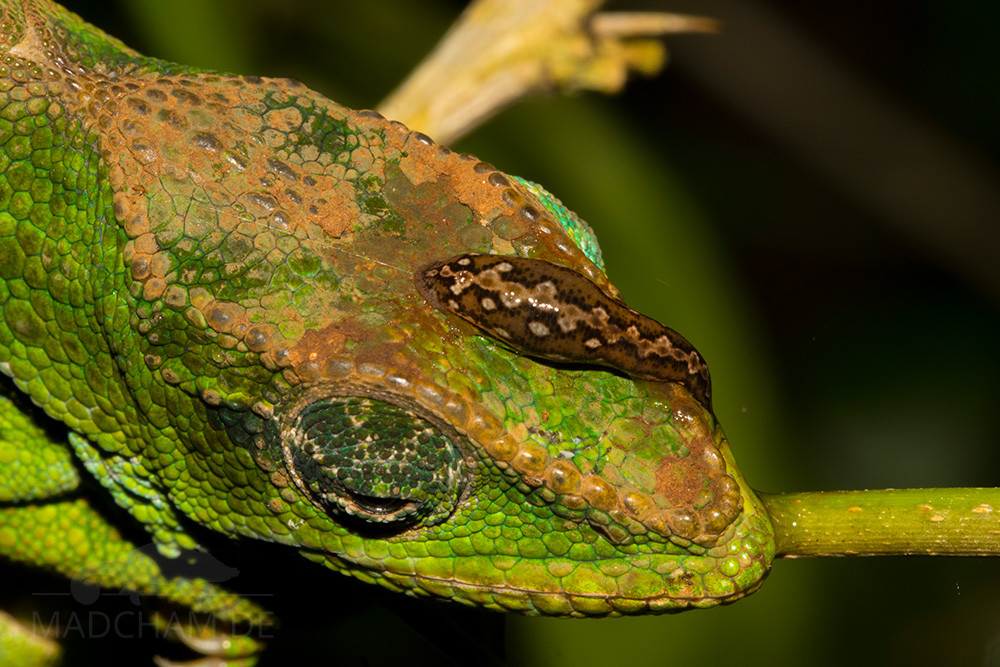
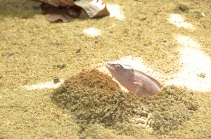
Calumma amber with a leech on its head in the Montagne d’Ambre
Leeches are common in Madagascar, but they appear to infest chameleons only very rarely. Unfortunately, studies on leeches that infest reptiles are rather scarce. There are some studies on the infestation of turtles and crocodiles by various species of leech. To date, there has been no research whatsoever into the occurrence of leeches on chameleons in Madagascar. We have so far observed a leech on a Calumma amber in the Montagne d’Ambre on one occasion. It was not possible to verify whether this was a coincidence or whether the leech was actually feeding on the chameleon. This therefore represents an exciting area for further research.
Das könnte dir auch gefallen


Follow us on Instagram


Most popular articles


Your travel specialist for Madagascar